Automated refraction test
A safe, high-precision infrared procedure for diagnosing sight problems (hypermetropia – myopia – astigmatism)
The procedure is used to establish eyesight levels in dioptres in order to make glasses or contact lenses, as prescribed by an eye doctor where necessary.
Using eye drops that temporarily inhibit the eye’s accommodation (medically induced cycloplegia) allows children’s eyesight to be determined with a greater degree of accuracy.
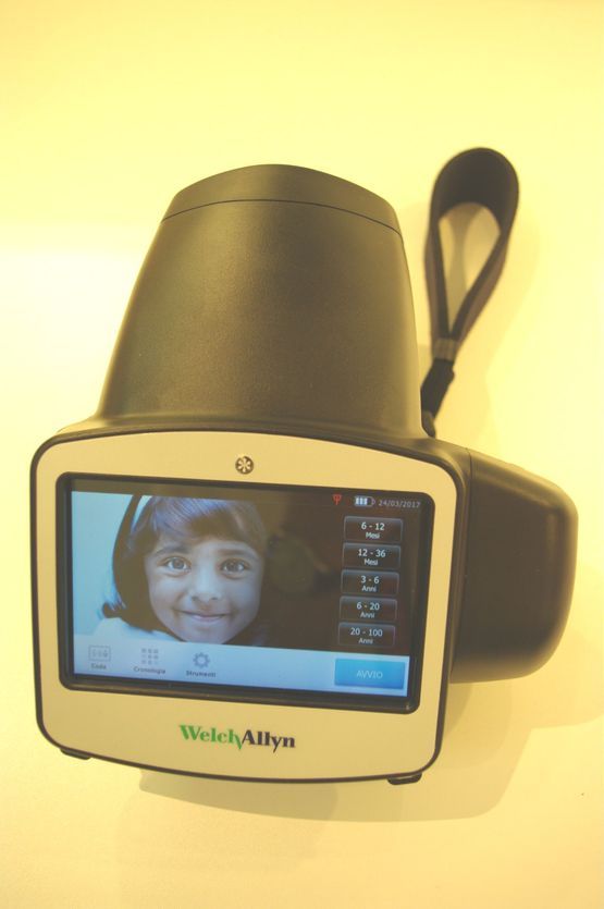
Spot vision screener (Welch Allyn)
This portable device enables the refraction test mentioned above to be carried out at a distance of one metre. It is most frequently used when examining small children (under four).
This method enables early diagnosis of sight problems and the creation of detailed reports for paediatricians.
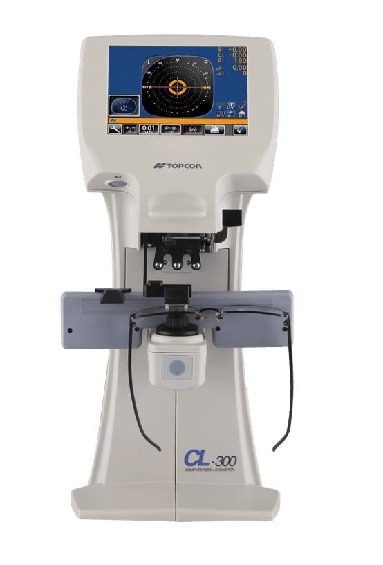
Verifying prescriptions (lensmeter)
A lensmeter establishes the refractive power of glasses (or contact lenses) in dioptres
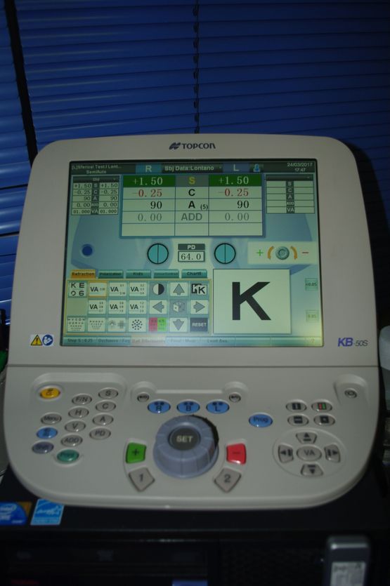
Sight test (phoropter)
This enables the computer-aided measurement of eyesight functioning and the correction of sight problems by prescribing visual aids.
Tonometry (measuring intraocular pressure)
Non-contact tonometry
: measurement using a short puff of air,
enabling rapid screening that can be carried out by a medical assistant.
Applanation tonometry following the application of anaesthetic eye drops:
- Slit lamp examination after applying anaesthetic eye drops
- using a Goldmann tonometer
- Tonometry using a resonance sensor:
- BioResonator®
- Tonometry using a hand-held Tono-Pen® device for children, bedridden patients and on house calls
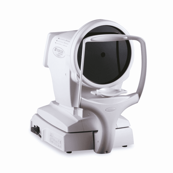
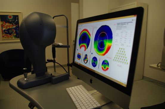
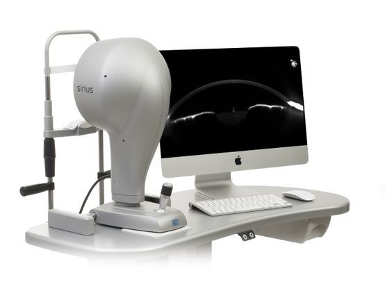
Corneal topography
Non-invasive examination of the quantity and quality of corneal tissue using videokeratography or a Scheimpflug camera with 360° rotation.
This makes it possible to measure the cornea’s refractive power and assess the front and rear corneal curvature.
The images also offer a topographical depiction of the corneal thickness (pachymetry).
Corneal topography enables early diagnosis of corneal surface and stromal conditions such as keratoconus.
This procedure is used for assessments before and after the surgical correction of sight problems using excimer lasers (PRK = photorefractive keratectomy or LASIK = laser-assisted in-situ keratomileusis).

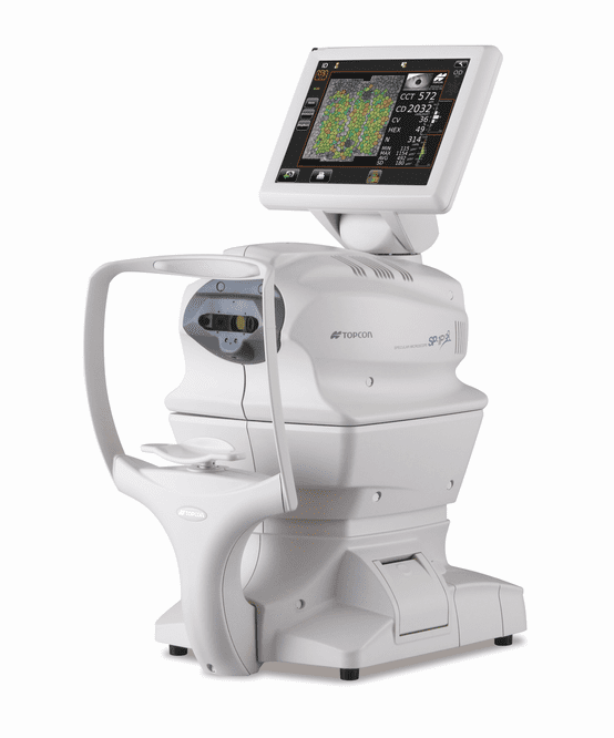
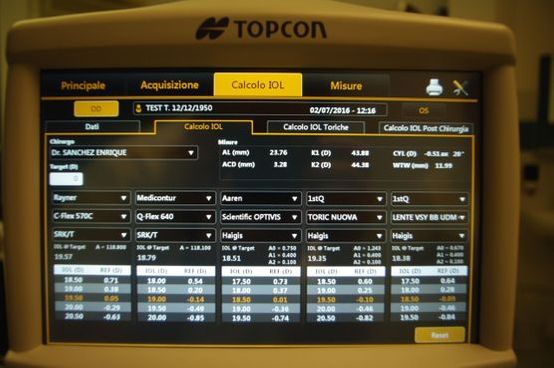
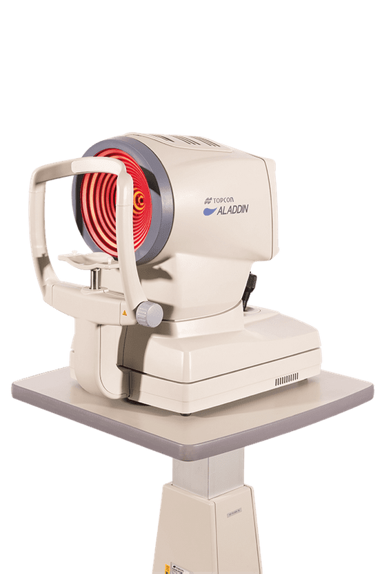
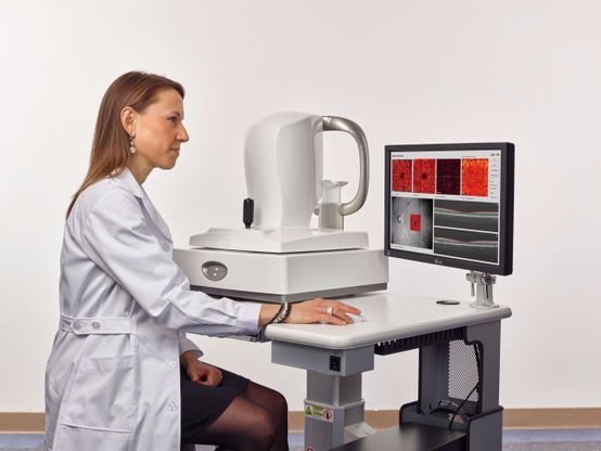
Biometry using an ultrasound probe
A biometric examination of the eye allows us to exactly measure its different parts.
The standard method uses eye drops followed by an ultrasound probe, and is non-contact, safe and painless (echography).
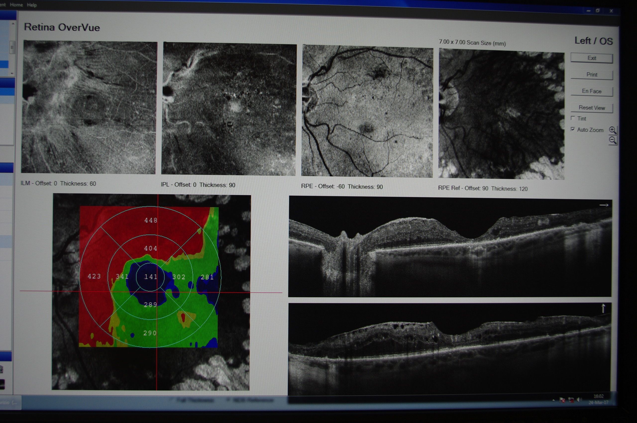
Optical coherence tomography (OCT)
OCT is used to diagnose various forms of macular degeneration, including dry and wet macular degeneration – the latter involving fluid leaking in or below the retina.
This procedure enables us to diagnose macular conditions earlier on and therefore offer more effective treatment.
OCT can also be used to diagnose corneal diseases and in aftercare for patients who have had laser treatment or have been treated for keratoconus using cross-linking.
OCT makes eye conditions easier to diagnose, such as:
- Macular oedema
- Diabetic retinopathy
- Age-related macular degeneration
- Central serous Retinopathy (inflammation of the choroid)
- Status following retinal vein occlusions
The procedure also allows us to monitor the effects of intravitreal injection of medication such as VEGF inhibitors (Lucentis, Eylea, Beovu, Avastin), corticosteroids (Ozurdex) and other drugs (e.g. Jetrea for the treatment of vitreomacular traction).
The procedure is completely safe and can be carried out on all patients.
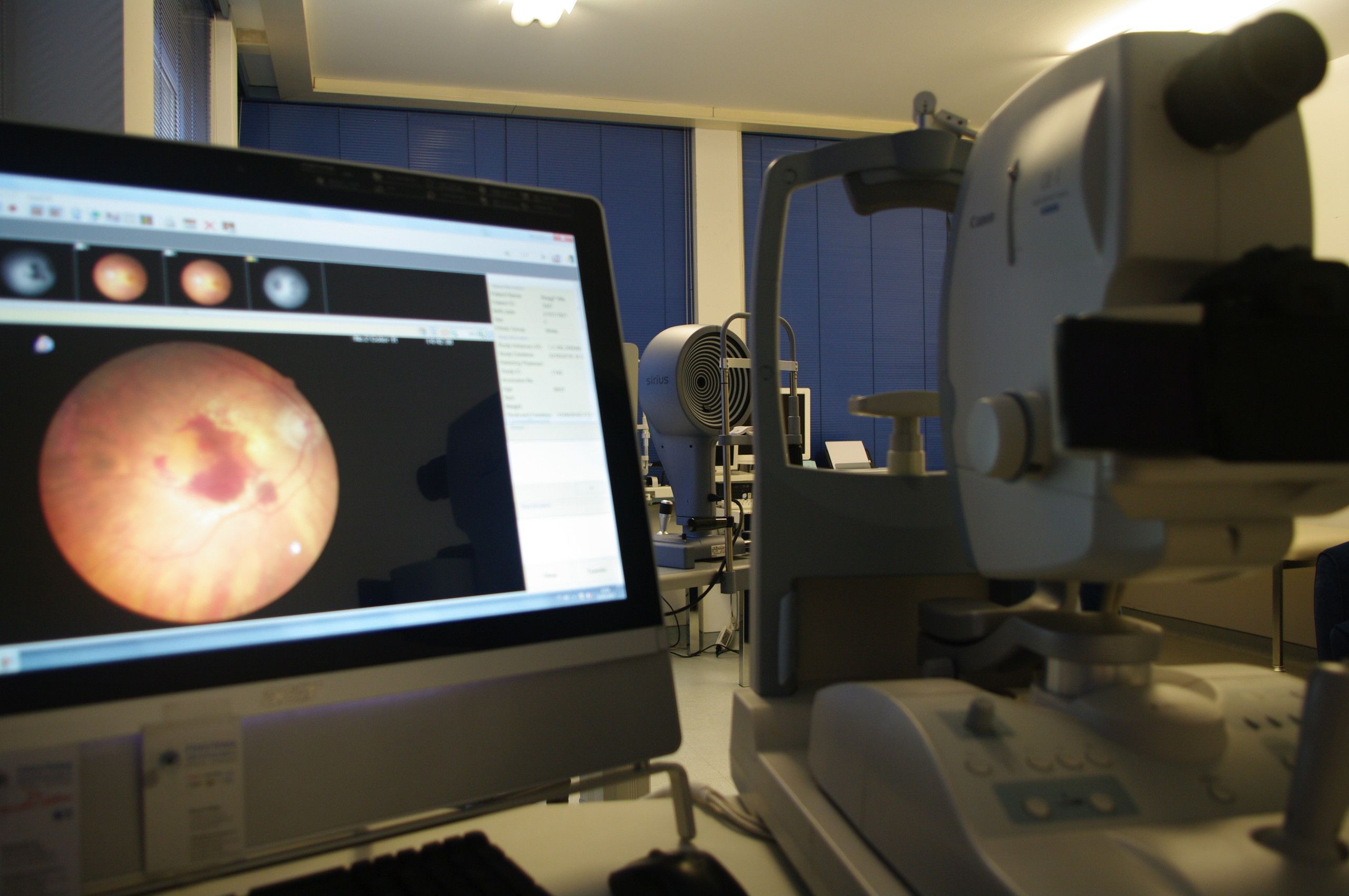
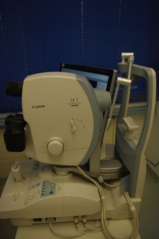
Fundus photography and angiography (fluorescein)
A digital retina camera enables:
- Colour images without dilatation (45°) or after dilatation, images of the retina (50°) with depictions of the retina, retinal circulation and the optic nerve
A special filter can be used for highly efficient assessment of nerve fibres (glaucoma).
Another filter enables autofluorescence imaging of the macula without using a dye, which is particularly relevant for macular diseases such as macular dystrophy and age-related macular degeneration.

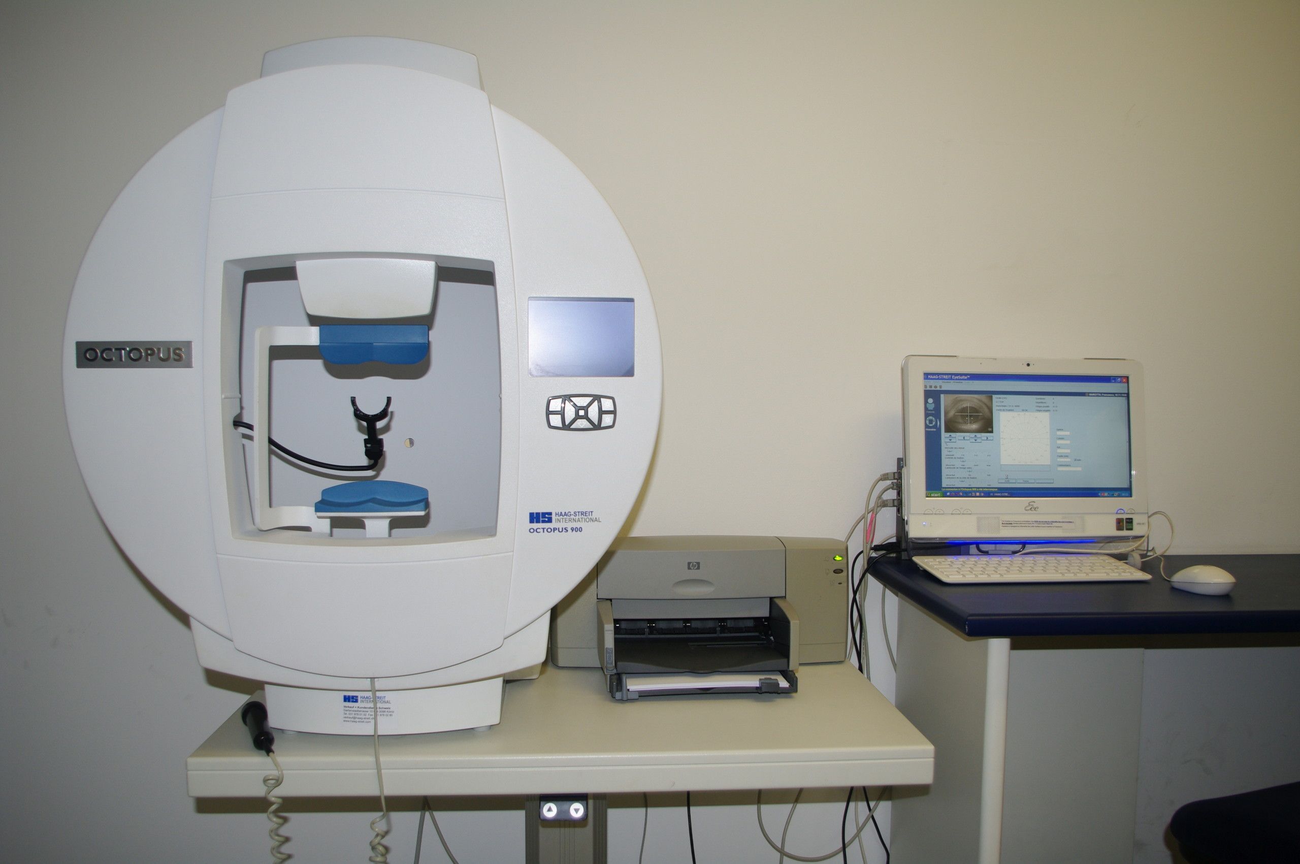
Perimetry
A) Kinetic perimetry:
this enables the field of vision to be measured using a light source that changes in size and intensity and moves from the periphery to the centre.
This fast, simple examination is used to identify neuro-ophthalmological diseases.
B) Computer-aided perimetry:
the patient focuses on a central point while flashes of light are emitted at different intensities.
This enables us to establish the threshold above which the light source is recognised, resulting in a graphic depiction of the field of vision for both eyes.
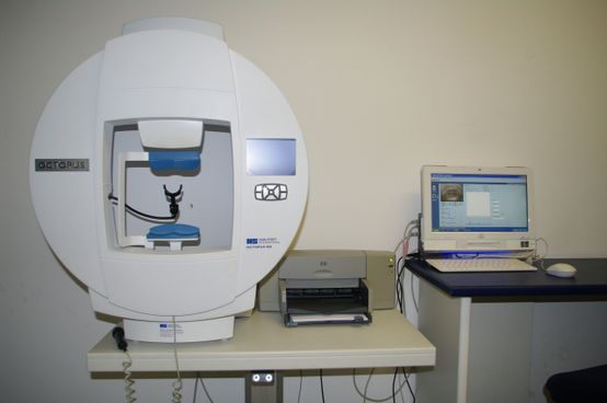
Octopus 600:
this enables an assessment of the central field of vision for both eyes with 60° rotation and includes Pulsar perimetry – a technology that measures both the contrast sensitivity and brightness of the stimuli, which is key for the early diagnosis of glaucoma.
Orthoptic examination using a Hess-Weiss graph (examining squints:
eye exercises using orthoptics)
Used for examining oculomotor dysfunction by projecting light onto a Hess-Weiss graph.
Used to diagnose oculomotor nerve palsy (neuro-ophthalmological examination), congenital
or trauma-induced strabismus (orbital fracture), vascular injury, diabetes or inflammation of the central
nervous system.
Colour test
- (24 tables)
- Farnsworth test (15 or 100 round colour plates)
Enables the diagnosis of congenital colour blindness, such as daltonism (red/green or yellow/blue colour blindness), or acquired vision problems such as macular or optic nerve diseases (often associated with inflammation or certain medications).
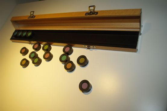
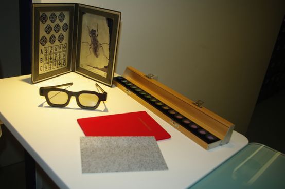
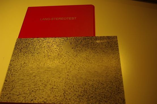
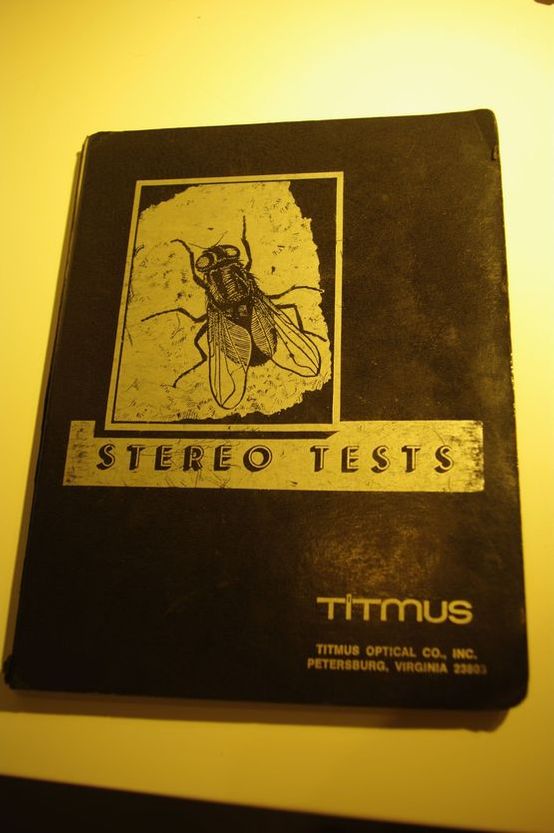
Stereoscopic sight test
Used to check three-dimensional vision using a simple test (Lang stereotest or the use of polarising filters).
This test is important for establishing fitness to drive.
Fitness to drive test (driving licence):
sight test with and without glasses, colour test, stereotest, eye motility and field of vision.
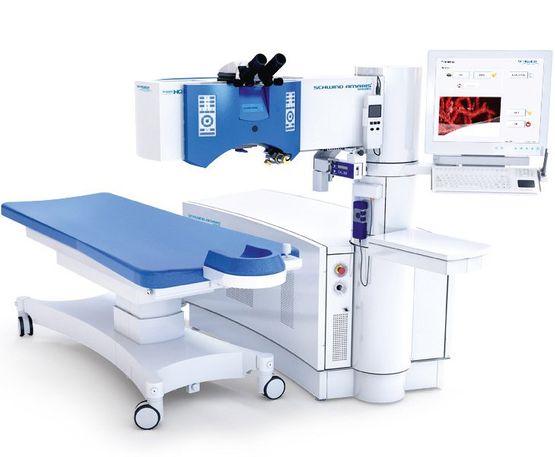
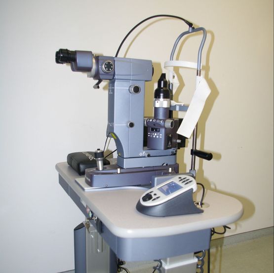
Laser treatments
When it comes to treating sight problems, opthalmologists have a wide range of treatments at their disposal. Laser treatments play a significant role.
The excimer laser can be used to correct corneal curvature and
scarring.
Patients can have their sight restored using a YAG laser following a capsulotomy or capsule cleaning (secondary cataracts).
For patients with angle-closure glaucoma, a peripheral iridotomy creates a bypass to improve the drainage between the anterior and posterior chambers while simultaneously reducing the intraocular pressure.
The laser is also highly effective for trabeculoplasty and trabecular cleansing.
The trabecular meshwork acts as a natural filter through which fluid from the chambers flows.
Patients with glaucoma usually experience a problem with their trabecular meshwork, which becomes “contaminated” with dead or inflamed cells or pigments which block the trabecula.
Selective laser trabeculoplasty (SLT) using a YAG laser does not harm the eye and can be repeated as many times as necessary.
Another method for treating glaucoma is the use of an external probe that allows selective access to the ciliary body, which contributes to the production of fluid in the eye chambers.
Cyclodestruction is carried out using a laser diode measuring 810 nanometres.
Another method is to create a barrier using an argon laser to combat holes in the retina, thereby preventing retinal detachment.
We make use of the following devices:
- Quantel Medical Argon 532-nanometre laser: for photocoagulation of the retina
- Quantel Medical YAG 1064-nanometre laser: for peripheral iridotomy and capsulotomy (secondary cataracts)
- Iridex Diode 810-nanometre laser: for photocoagulation of the retina and tumour treatment using transpupillary thermotherapy (TTT), which uses an external probe to carry out cyclodestruction to combat neovascular glaucoma and other forms of treatment-resistant glaucoma
- Amaris 500 excimer laser: a high-frequency, 500-Hertz laser used to correct all varieties of sight problem (hypermetropia, astigmatism, myopia), and for combined treatments such as for presbyopia using PresbyLASIK
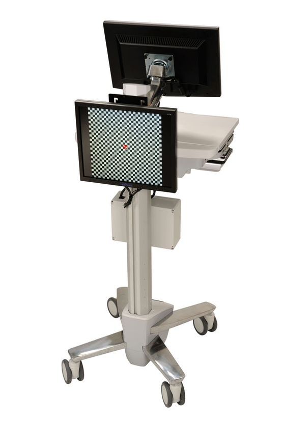
Diopsys electrophysiology
Diopsys ERG is a non-invasive test measuring the electrical functionality of the retina, in particular the macular and ganglion cells.
There are two ERG test methods available,
one using concentric stimuli and one using contrast sensitivity.
The test is swift and simple, and can be repeated as necessary.
An adhesive pad with recording electrodes is attached to the skin, and the patient looks at a monitor which displays visual stimuli.
The test takes just a few minutes, after which the electrodes are removed and an ERG report is immediately available.
Diopsys ERG is used to diagnose and treat retinal diseases such as age-related macular degeneration (AMD), diabetic macular oedema, diabetic retinopathy and toxic maculopathies (such as
maculopathy related to taking Plaquenil).
It enables us to assess the functionality of ganglion cells in the retina, allowing us to diagnose glaucoma at an early stage.
It helps doctors assess the results of a selected treatment method, e.g. use of medication (eye drops) to reduce intraocular pressure, selective laser trabeculoplasty or surgery.
In conjunction with other tests such as computer-aided assessment of the field of vision and anatomical examinations of the optic nerve and nerve fibres using OCT, Diopsys ERG can be used to assess and monitor every eye patient.