Diopsys electrophysiology
Diopsys ERG is a non-invasive examination measuring the electrical functionality of the retina, in particular the macula and ganglion cells.
There are two ERG test methods available, one using concentric stimuli and one using contrast sensitivity.
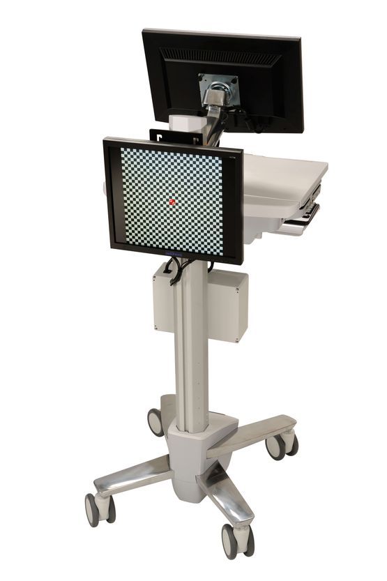
It is a swift, simple, safe examination that can be repeated as necessary. The first step is attaching adhesive recording electrodes to the skin.
The patient then looks into a monitor that displays visual stimuli.
The examination only takes a few minutes.
The results of the ERG are available immediately once the electrodes have been removed.
Diopsys ERG is a helpful tool for diagnosing and carrying out follow-up examinations of retinal diseases such as aged-related macular degeneration (AMD), diabetic macular oedema, diabetic retinopathy and toxic maculopathy (e.g. maculopathy related to taking Plaquenil).
It enables an evaluation of the functionality of the retinal ganglion cells and thus an early diagnosis of glaucoma. It also helps doctors assess the results of a selected treatment method, e.g. use of medication (eye drops) to reduce intraocular pressure, and following selective laser trabeculoplasty or surgery.
Diopsys ERG can be used to assess and follow up with every eye patient in conjunction with other tests such as computer-aided assessment of the field of vision and anatomical examinations of the optic nerve and nerve fibres.
Keratoconus screening
What is keratoconus?
Keratoconus is a degenerative disease affecting the cornea in which the corneal tissue progressively thins and distorts, leading to an uneven astigmatism, often in conjunction with short-sightedness.
There is a genetic element to developing this disease. Usually both eyes are affected.
There are also behavioural risk factors, such as rubbing your eyes very hard on a regular basis.
Diagnosing keratoconus:
Keratoconus can be diagnosed as early as puberty, or in young adults. The progression of the disease slows after the age of 40.
Specialist centres can diagnose the condition by measuring the corneal thickness and the anterior and posterior corneal curvature, and by carrying out a corneal topography, i.e. creating a “map” of the cornea.
Treatment:
sight problems can only be treated in the early stages using glasses.
For advanced corneal curvature, semi-rigid contact lenses are usually necessary.
Until recently, the standard surgical treatment was a cornea transplant, which had excellent results in terms of vision but came with a significant risk of post-operative transplant rejection, and therefore required the patient to take corticosteroids and other immunosuppressants such as interferons, and to go for regular check-ups with their eye doctor.
- Cross-linking (CXL):
- this is a safe, simple and effective method of slowing the progression of keratoconus.
- Rings or ring segments made of transparent plastic (PMMA):
- these are implanted into the cornea to limit the protrusion of the centre of the cornea and the astigmatism, thereby improving the patient’s eyesight.
What happens during cross-linking (CXL)?
Cross-linking is a non-invasive procedure that attempts to stabilise the structure of the cornea using the combined effects of riboflavin (vitamin B2) and UVA radiation in order to create new, stronger connections or “links”.
There are two ways of carrying out the procedure:
- The traditional approach removes the epithelium (“epithelium-off”) before cross-linking (CXL)
- Transepithelial cross-linking is a newer approach that does not remove the epithelium (“epithelium-on”) before cross-linking (CXL).
- The second option, transepithelial cross-linking (CXL), has a number of advantages for the patient, both in terms of safety and comfort and with regard to post-operative sight restoration.
- Refraining from removing the epithelium makes the procedure painless and reduces the risk of infection to almost zero.
- Patients can return to work immediately after the procedure.